Getting diagnosed with melanoma
Many people start by seeing their GP if they have symptoms that may be due to melanoma skin cancer. Your GP can do some tests to help them decide whether you need to be referred to a specialist. They might:
do a physical examination of your skin
look closely at your mole or skin with a special instrument (dermatoscope)
take or arrange photographs of the mole or abnormal patch of skin
Your GP will look at the mole or abnormal area of skin. They may measure it with a ruler or a special marker scale. They can compare the size, shape and colour to other moles you have or other areas of your skin.
Your GP may also feel around your neck, armpits or groin. This is to see if your are swollen.
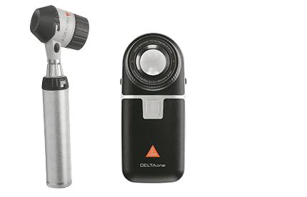
Your GP might look at your mole or skin through a special instrument called a dermatoscope. It makes things look bigger, like a magnifying glass.
Below are examples of dermatoscopes.

Your GP puts some gel or oil on the mole or abnormal area of skin. They put the dermatoscope on top and look through it. This lets them examine the area very closely. The oil or gel helps the dermatoscope work better. It does not hurt or damage the skin.
Some GPs are trained to use a dermatoscope. If your GP isn't, a skin specialist will look using a dermatoscope, if you need to see them.
Read more about having a dermoscopy
Sometimes your GP might want a specialist skin doctor (dermatologist) to look at the mole or abnormal area of skin. This is to help them decide if you need to be referred to the hospital. Your GP will take some photos of the mole or abnormal area of skin to send to the dermatologist. This is called teledermatology. If your GP can’t take the photographs you might have them taken at a special clinic.
Your GP may ask you to take photographs of the mole or skin patch. They will tell you the best way to take them. They will also tell you how to safely send the photographs.
Depending on the features of the abnormal mole or area of skin your GP may refer you to a specialist doctor. You usually see a dermatologist. Sometimes you might see a .
The specialist doctor usually arranges more tests. These may include:
dermoscopy if not done by your GP
taking photographs to see if the mole or abnormal patch of skin changes (baseline photographs)
removing the mole or abnormal patch of skin (excision biopsy) to find out if you have melanoma
Your doctor uses photographs to keep a record of what the mole or area of skin looked like when you first saw them. These are called baseline photographs.
Sometimes they will ask you to come back in a few months to see if the mole or patch of skin has changed. They do this by comparing it to the baseline photographs. They will then decide if it needs to be removed or not.
Your doctor might remove the abnormal area to find out if it's a melanoma skin cancer. This is called an excision biopsy. You have this under a so you will be awake during the procedure.
You lay on a couch and your doctor puts an injection of local anaesthetic into the skin around the mole or abnormal area. This makes the area numb. They remove the abnormal area and a small amount of skin (around 2mm) around it. This is called the margin.
They send the abnormal area or mole to the laboratory. A specialist doctor (pathologist) looks at the tissue under a microscope. This is the only way to know for certain if you have melanoma.
Your doctor might use stitches to close the wound. These might dissolve on their own or a nurse may need to remove them. You may have a small dressing over the top. Your doctor or nurse will tell you:
if the stitches need to be removed
where and when to get the stitches removed
how to look after the wound and dressing
You usually go home the same day. Your doctor will tell you when to expect the results. It can take a couple of weeks.
Your doctor will normally recommend an operation to remove a larger area of skin around where the melanoma was. This is called a wide local excision.
They might also recommend you have tests:
to check your lymph nodes
on the melanoma to look for changes (mutations)
If you have a wide local excision for an early stage melanoma you may not need any more tests afterwards. Your doctor will talk to you about this.
Read more about having surgery to remove melanoma skin cancer
Lymph nodes are glands. They are found in many parts of the body including the armpits, neck and groin. They help drain away waste fluid and damaged cells. They also contain cells that fight infection.
There is a risk that melanoma cells can spread to the lymph nodes. The risk is higher in thicker melanomas. But it’s rare in melanomas that are less than 1mm thick.
Your doctor may check your lymph nodes for melanoma by taking a biopsy. Which lymph nodes they take the biopsy from depends where on your body the melanoma skin cancer was.
There are different types of lymph node biopsy. Which one you have depends on:
the size of the melanoma
if your doctor can feel your lymph nodes (palpable lymph nodes)
how easy it is to take a biopsy of the lymph node
The first place that melanoma skin cancer usually spreads to is the nearby lymph nodes. The sentinel lymph nodes are the first lymph nodes the cancer can reach.
During an SLNB your doctor removes the sentinel nodes and sends them to the laboratory. This is to check them for small amounts of melanoma that can only be seen under a microscope (microscopic disease).
You have an SLNB under a at the same time as the wide local excision.
Read more about having a sentinel lymph node biopsy for melanoma skin cancer
Sometimes your doctor might recommend a different type of lymph node biopsy. This is either a fine needle aspiration (FNA) or core needle biopsy. You usually have one of these if your doctor can feel your lymph nodes are swollen, or they look swollen on an ultrasound scan.
Your doctor can use an ultrasound scanner to help them take the biopsy. They send the sample of fluid or tissue to the laboratory to be checked for cancer cells.
Your doctor uses a thin needle to go through your skin and into the swollen lymph node. They use a syringe at the end of the needle to take out some fluid and small pieces of tissue.
This is similar to an FNA but your doctor uses a thicker needle. This means they can remove a larger amount of tissue.
Your doctor injects some local anaesthetic around the swollen lymph node before you have a core needle biopsy. This makes the area numb.
Read more about having a lymph node ultrasound and biopsy
For stage 3 and 4, and some stage 2 melanoma skin cancers, your doctor tests the melanoma cells for gene changes (mutations). This includes the BRAF V600 gene. They usually do the genetic tests on the melanoma that was removed. But sometimes they need to take another sample. For example, from where the cancer has spread.
Changes in the BRAF V600 gene cause the melanoma to make lots of BRAF protein. This protein tells the melanoma cells to grow and divide. Having lots of the protein means the cells grow and divide more quickly.
If you have a change in the BRAF V600 gene, doctors describe the melanoma as BRAF positive or BRAF mutant. If you don’t have the change, then the melanoma is called BRAF negative or BRAF wild type.
Knowing if you have a gene change helps your doctor recommend the most suitable treatment for you.
Read more about treatment for melanoma skin cancer
Doctors use CT or MRI scans to check if the melanoma has spread. Not everyone will need these.
CT (or CAT) scan stands for computed (axial) tomography. It is a test that uses x-rays and a computer to create detailed pictures of inside your body.
You may have to drink a jug of water or contrast medium before the test. This is a dye that shows up body tissues more clearly on the scan. You might also have an injection of contrast medium.
Read more about having a CT scan
MRI stands for magnetic resonance imaging. It uses magnetism and radio waves to take pictures of inside your body.
Read more about having an MRI scan
Your doctor usually wants you to have a CT or MRI scan if:
a biopsy shows melanoma in your lymph nodes
you don’t have melanoma in your lymph nodes but your doctor thinks the melanoma has spread
you haven’t had a lymph node biopsy but your doctor thinks the melanoma has already spread
You usually have a scan that looks at your:
chest
tummy (abdomen) – including the area between your hips, called the pelvis
head
If your doctor thinks you should have MRI scans instead of CT scans they will talk to you about this. Some people may have a CT scan of their chest, abdomen and pelvis and an MRI scan of their head.
A blood test is where your doctor, nurse or takes a small amount of blood and sends it to the laboratory. Lots of changes in your body show up in blood tests.
You may have blood tests to check your general health, including how well your kidneys and liver are working. Your doctor may also check the number of blood cells. You usually have blood tests if you are having:
an operation under general anaesthetic to remove melanoma skin cancer
a CT or scan – this is to check your kidneys are working well and are able to get rid of the special dye (contrast) they use for the scan
targeted cancer drugs, immunotherapy or chemotherapy
You might have a blood test to check your vitamin D levels. Your skin produces vitamin D when you have been in the sun. It is needed for healthy bones.
Doctors ask people with melanoma skin cancer to protect their skin from the sun. This means they may have lower levels of vitamin D in their body. You can also get vitamin D by eating eggs and oily fish such as mackerel and salmon. But if your Vitamin D level is low, your doctor may ask you to take a vitamin D tablet (supplement).
Read more about having a blood test
Occasionally your doctor may want you to have other tests. This might include a PET-CT scan. This is a combination of a PET scan and a CT scan.
Your doctor will explain what these tests are for and how to prepare for them.
Find out about having a PET-CT scan and other tests
The tests you have help your doctor find out if you have melanoma skin cancer and if it has spread. This is called the stage of a cancer. This is important because doctors recommend your treatment according to the stage.
Find out about the stages of melanoma and the treatment
Coping with a diagnosis of melanoma skin cancer can be difficult. There is help and support available for you and your family.
Last reviewed: 02 Jan 2025
Next review due: 02 Jan 2028
Melanoma skin cancer starts in skin cells called melanocytes. You can get it anywhere on your skin including in a mole, on your palms, the soles of your feet and under your nails.
Symptoms include changes to a mole, freckle or normal patch of skin. Doctors use a checklist of signs to look out for. But it helps to know what your skin normally looks like.
See your GP if you develop a new mole, abnormal area of skin or changes to an existing mole. They will look at it and may refer you to a specialist.
You may be referred to a specialist if you have symptoms that could be due to melanoma skin cancer. This might be an urgent suspected cancer referral.
Find out about tests to diagnose cancer and monitor it during and after treatment, including what each test can show, how you have it and how to prepare.
Melanoma develops in cells called melanocytes. You have these in your skin and other parts of your body. Melanoma that starts in the skin is called melanoma skin cancer.

About Cancer generously supported by Dangoor Education since 2010. Learn more about Dangoor Education
Search our clinical trials database for all cancer trials and studies recruiting in the UK.
Connect with other people affected by cancer and share your experiences.
Questions about cancer? Call freephone 0808 800 40 40 from 9 to 5 - Monday to Friday. Alternatively, you can email us.